Introduction
ScanImage allows the microscope's full field of view to be subdivide into multiple regions of interest (ROIs).
The use of ROIs offers the following advantages:
- Frame Rate Optimization
- Limiting tissue exposure
- Tracing features through a volume
Frame Rate Optimization
In a traditional laser scanning microscope, the frame rate is calculated as follows:
frame rate [Hz] = 1 / (lines per frame * line period [s])
To increase the frame rate, the line period and/or the number of lines per frame can be decreased. Since the number of lines per frame directly translates to the vertical resolution of the image and the line period is either hardware constrained (resonant scanning) or would result in a decreased horizontal resolution (galvometric scanning), this method is often unacceptable for scenarios where a high image quality is desirable. For small objects that are distributed over a large range of the microscope's field of view, a compromise between frame rate and detail resolution needs to be found.
To overcome this limitation, ScanImage supports the definition of regions of interest (ROIs). ROIs allow to optimize the frame period by skipping image regions of low interest, while preserving the image resolution within the ROI.
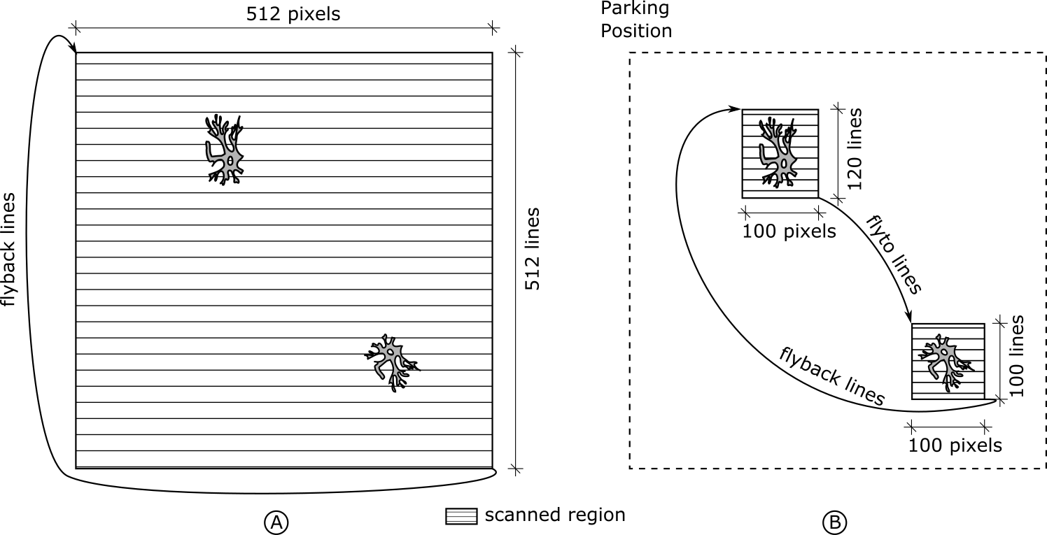
A: When scanning the full field of view, regions of low interest contribute to the frame period and therefore decrease the frame rate.
B: By defining regions of interests (ROIs) the number of scanned lines is reduced to maximize the framerate while preserving the image resolution within the ROIs
Limiting Tissue Exposure
ScanImage allows to control the laser power by using Pockels cell in the optical path. By fully attenuating the beam power while traversing between ROIs, the exposure of tissue is minimized. Additionally, ScanImage allows beam power to be set differently for each ROI. This allow you to limit power differently to certain regions if some are more sensitive than others.
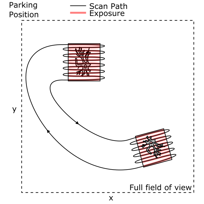
Tissue is only exposed to the laser while image data is acquired.
The beam is attenuated in between regions of interest.
Tracing of features through a volume
A ScanImage volume is the collection of multiple image planes at different z-planes. Typically, the volume is scanned by using a piezo actuator to axially sweep the microscope's objective while continuously acquiring images. This way each acquired frame represents one slice of the volume.
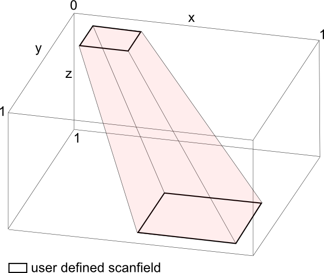
In ScanImage's multi-ROI feature, a ROI is the generalization of a standard volume. To define a ROI a user can define 'scanfields' at multiple cross sections. A scanfield is defined by x,y position, x,y extent, rotation and resolution. It is sufficient to define scanfields at a ROI's top and bottom cross sections since ScanImage interpolates scanfields within the volume as needed.
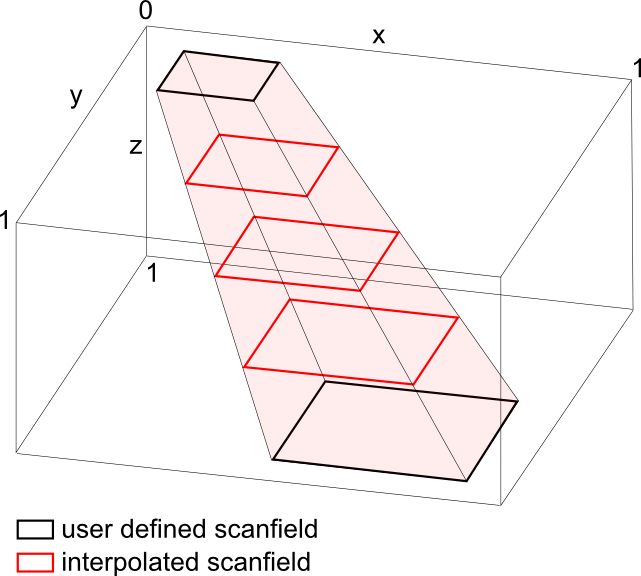
By defining multiple scanfields at different z-planes of a ROI, features can be traced through a volume.
Defining ROIs to Image
A ROI Group consists of a collection of ROIs to be imaged. Each ROI is defined by a set of scanfields that define the ROI's cross section at a particular Z plane. The following articles describes ROI Groups, ROIs, and scanfields in more detail, and how to use the ROI Group Editor to create ROIs.